AI Microscope Captures 'Family Portrait' of 15 Cell Structures
Eastern Institute of Technology, Ningbo's Assistant Professor Karl Zhanghao and collaborators integrate AI and 'optical fingerprinting' to achieve simultaneous dynamic observation of 15 subcellular structures in living cells for the first time, breaking through the channel number limit of traditional cell panoramic imaging. The related research results were recently published in Nature Communications.
AI Microscope Achieves Synchronous Dynamic Observation of 15 Subcellular Structures
If living cells are compared to a precisely operating micro-factory, various organelles within the cell are functional workshops with specific responsibilities, while microscopic imaging acts like a real-time monitoring system. Traditional multicolor imaging is limited by spectral interference and phototoxicity, making it difficult to simultaneously observe the collaborative processes of more than 6 types of organelles. Moreover, traditional multicolor imaging typically uses a time-sharing approach, so more imaging channels also face lower temporal resolution. This technical bottleneck where "high speed" and "multiple types" cannot be achieved simultaneously has long hindered research on the complete interaction network of organelle dynamics.
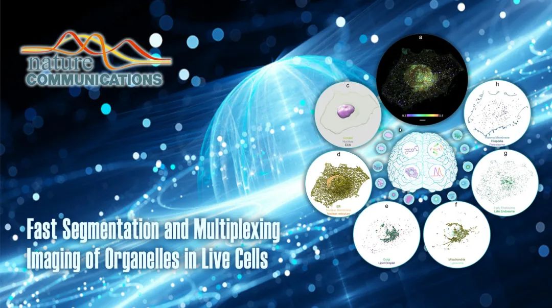
The research team used the lipophilic probe Nile Red to uniformly stain all membrane-structured organelles, capturing the unique spectral ratio "fingerprints" of different organelles with a spinning disk confocal microscope. They then constructed an Attention U-Net deep neural network, using super-resolution fluorescence images and spectral ratio images as dual input channels, combined with ground truth data from organelle-specific markers to train the model, ultimately establishing an AI system that can accurately segment 15 subcellular structures.
This method successfully achieved dynamic imaging of mitochondria, endoplasmic reticulum, lipid droplets, cell membrane, lysosomes, endosomes, Golgi apparatus, nuclear envelope, peroxisomes, cellular pseudopods, nuclear invaginations, as well as the nucleus, cytoplasm, and extracellular matrix, laying a technical foundation for mapping the complete organelle interaction atlas.
