Spectral-Polarization Optical Tomography: Building a Multi-Dimensional Super-Resolution "Street View Map" of Organelle Interactions
Spectral-Polarization Optical Tomography: Building a Multi-Dimensional Super-Resolution "Street View Map" of Organelle Interactions
Just like a city, a cell is a fully functional "microscopic world" containing various organelles responsible for material transport, metabolism, genetic inheritance, and endocrine regulation. Research on organelle interactions is crucial for understanding cell functions and the root causes of diseases. However, current fluorescence microscopy is limited by factors such as finite fluorescence color channels, staining varieties, dye stability, and temporal-spatial imaging resolution.
Lipid membranes widely exist in subcellular organelles, whose morphology, composition, and lipid phases cooperatively regulate biophysical membrane properties, membrane protein functions, and interactions between lipids and proteins. Although lipid membranes play important roles in subcellular organelle biochemical functions and interactions, their classification, interaction research, and long-term dynamic observation are challenging due to their similar chemical compositions.
Recently, the research team developed Spectral-Polarization Optical Tomography (SPOT) technology. Combined with lipophilic probes, SPOT simultaneously images lipid membranes of 10 subcellular organelles and analyzes their lipid dynamics from three optical dimensions: intensity, spectrum, and polarization. This work was recently published in Nature Communications.
Compared with existing fluorescence imaging technologies, this independently developed SPOT technology obtains information from multiple dimensions including fluorescence intensity, spectrum, and polarization using only six original images. The imaging speed enables real-time observation of subcellular organelle dynamics. The technology's excellent optical sectioning capability simultaneously improves polarization detection accuracy and spectral detection precision, achieving for the first time lipid heterogeneity dynamics within subcellular organelles using optical imaging technology, enabling quantitative observation of lipid polarity and phase.
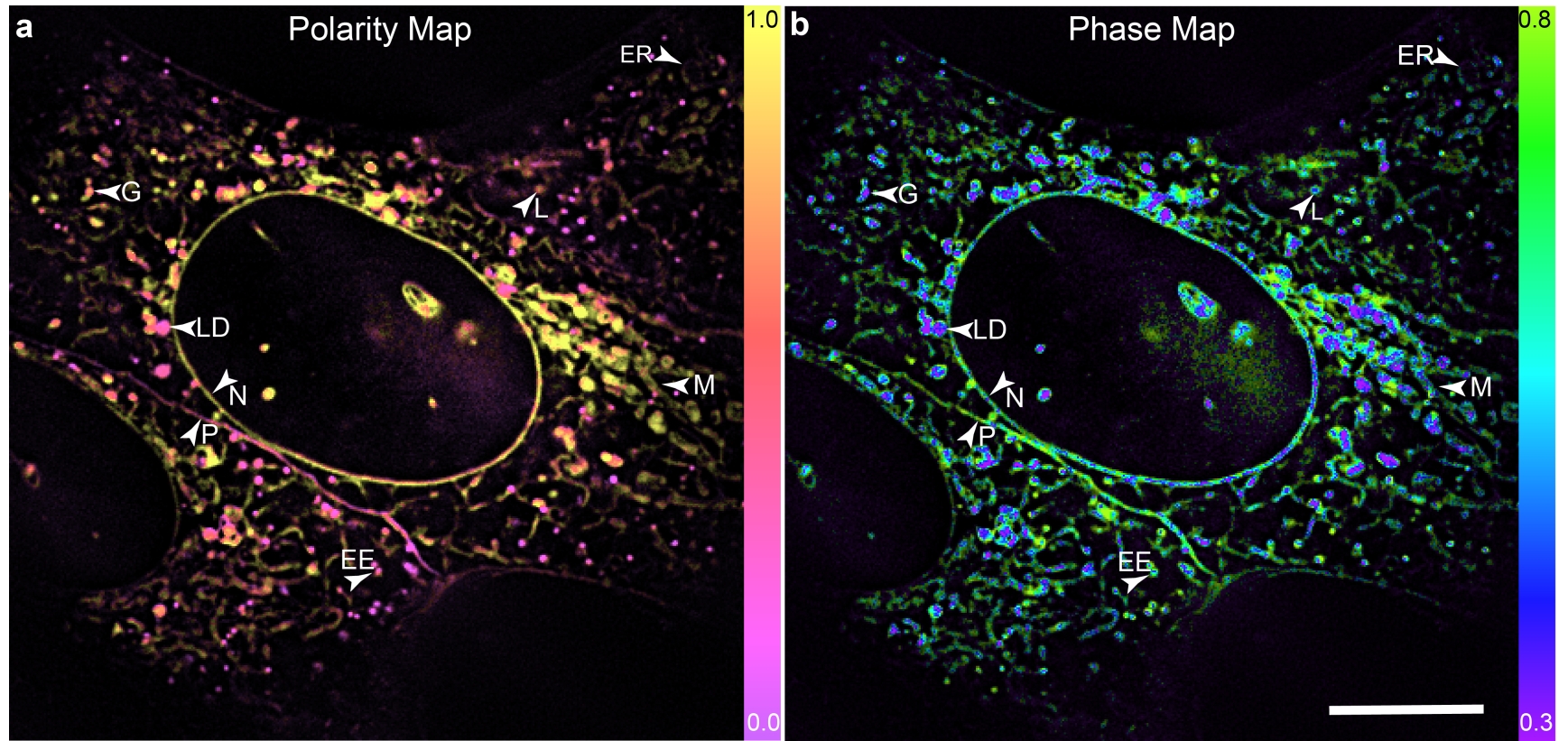
Figure 1 SPOT achieves lipid membrane heterogeneity analysis in subcellular organelles
Using SPOT technology, researchers discovered lipid heterogeneity in mitochondrial inner ridges and outer membranes, as well as lipid composition changes in endosomes during maturation. Through real-time monitoring, researchers captured dynamic changes in cell membrane heterogeneity during cell division, and dynamic changes in lipid composition during TNT formation and mitochondrial ridge disappearance processes.
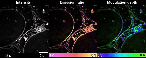
Figure 2 SPOT monitors dynamic changes of lipid membranes during cell division
Traditional fluorescence microscopy is limited by labeling methods, allowing simultaneous imaging of only up to four organelles. SPOT breaks through traditional optical imaging dimension limitations, achieving high spatiotemporal resolution live-cell imaging from six dimensions: three-dimensional space, time, polarization, and spectrum. Through the coordination of membrane morphology, lipid polarity, and lipid phase, SPOT enables simultaneous imaging and classification of ten subcellular organelles, adding a powerful tool for lipidomics and organelle interaction research.
Dr. Karl Zhanghao, Research Assistant Professor at Southern University of Science and Technology, Wen-Liu Liu, PhD student at Tsinghua University, and Mei-Qi Li, PhD student at Peking University, are co-first authors of this work. Prof. Peng Xi from Peking University College of Engineering, Prof. Dayong Jin from Southern University of Science and Technology, and Research Assistant Prof. Karl Zhanghao are co-corresponding authors. This work was supported by the National Natural Science Foundation of China, the Ministry of Science and Technology Key R&D Program, the Beijing Natural Science Foundation, and the Shenzhen Science and Technology Innovation Commission.
Original Article: https://doi.org/10.1038/s41467-020-19747-0
